In vivo
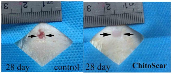
Figure 1 Comparison of wounds diameter on day 28 in experimental groups
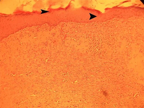
Figure 2 Histopathological section of skin in ChitoScar group in Day 28, complete epithelialization with keratinization (arrow head) is seen (H&E×10)
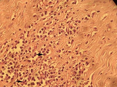
Figure 3 Histopathological section of skin in ChitoScar group in Day 28, marked leukocyte infiltration (arrow) is seen
(H&E×40)